Currently, I'm working on the visualization and quantification of (trace) element distributions in biological cells using X-ray induced X-ray fluorescence (XRF), at the sector 2 (2-ID-E/D) XRF microprobes of the Advanced Photon Source.
A somewhat 'typical' layout for a X-ray fluorescence microprobe is as follows: the pink undulator beam is monochromatized, e.g., by using a double crystal monochromtator. A zone plate objective is used to focus X-rays onto the specimen, an order sorting aperture rejects unfocused X-rays to reduce the background.
The sample is raster scanned through the focal spot, and at each scan position, illuminated with X-rays.
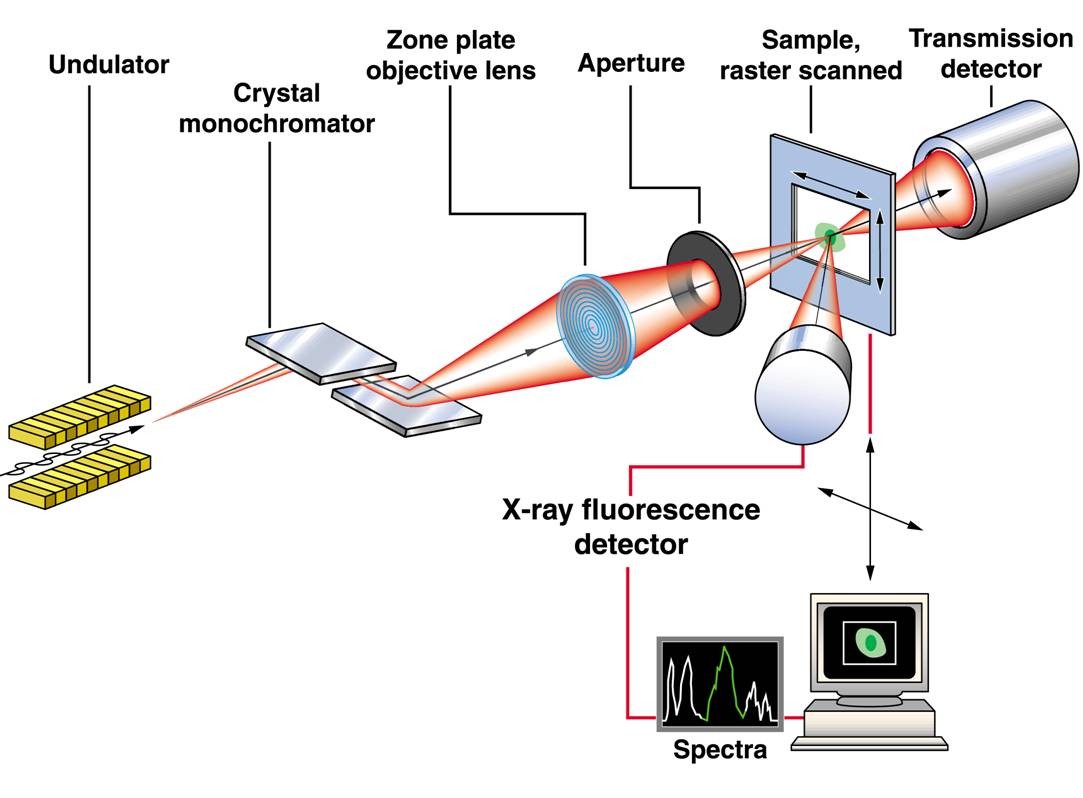
The incident X-rays excite photo electrons in the the sample. The vacancies in the inner shells of the atoms that result are filled in by outer shell electrons, either through an Auger process where the excess energy between inner and outer shell electron binding energies is carried away by a secondary electron, or through a fluorescence process where the excess energy is carried away by a photon. In X-ray fluorescence mapping, the latter can be detected using an energy dispersive detector system. By measuring the amount of electron hole pairs generated by the fluorescence photon, the chemical element from which it originated can be deduced. Since the number of detected fluorescence photons goes linear with the quantity of material present in the illuminated spot on the sample, the amount of material can be quantified by using proper elemental standards. Typically, we map (and quantify) 10 or more elements simultaneously at each scan position.